Applied laboratory science never ceases to amaze me!
Take Van Gieson's picro-fuschine staining for collagen fibers. In this solution, you have two anionic dyes, picric acid and fuschin acid, which theoretically stain the same cationic structures. So how is it that the staining gives us a clear and easily distinguishable red collagen stained with fuschin and the other structures (cytoplasms and globules) stained yellow with picric acid?
It would seem that the difference in size of the two molecules is the key to understanding this enigma. Smaller picric acid molecules are more likely to bind to dense structures, while larger molecules like fuschin are more likely to stain loose tissues like collagen. Picric acid's rapid diffusion would also contribute to this effect, staining quickly whereas fuschin acts more slowly. And once the chromophores are attached, salt bonds are created, contributing to the dye's diffusion by absorption.
The result depends more on the porosity and permeability of the different tissues to be stained than on competition between two anionic chromophores! Surprising all the same! The same principle also applies to Masson's Trichrome between Briebrich scarlet and fuschin. Of course, as always when working with picric acid, one has to be extremely careful because, in addition to being toxic and staining skin and clothes for a long time, this product is explosive when dry (or dried on the neck of the bottle!) Ready-to-use solutions are available on the market to avoid these worries.
All these physical principles have been used in histology for a long time and are still in use today! Thank you Mr. Ira Van Gieson!
1. What is the purpose of the Van Gieson picric acid–acid fuchsin stain?
It is used to highlight collagen fibers, clearly distinguishing them from other tissue structures.
2. What are the two dyes used in this stain?
The two dyes are:Picric acid (anionic dye)
Acid fuchsin (also an anionic dye)
3. Why is it surprising that these two dyes produce different colors in tissues?
Because both dyes are anionic and would theoretically stain the same cationic structures. Yet, they stain different tissues in distinct ways.
4. What property mainly explains the different staining patterns between these two dyes?
The size of the molecules:The small picric acid molecules penetrate and bind better in dense structures (cytoplasm, red blood cells).
The larger acid fuchsin molecules stain looser tissues such as collagen.
5. What role does diffusion speed play in the final result?
Picric acid diffuses quickly and stains dense tissues rapidly.
Acid fuchsin diffuses more slowly, allowing it to preferentially bind to collagen.
6. Is the result due to chemical competition between the two dyes?
No. The result depends more on tissue porosity and permeability than on competition between the dyes.
7. What other staining protocol works according to the same principle?
The Masson Trichrome stain, where Biebrich scarlet and acid fuchsin distribute according to tissue density.
8. What precautions must be taken with picric acid?
It is toxic.
It can permanently stain skin and clothing.
It is explosive when dry, including when it dries on the bottle cap.
Therefore, ready-to-use solutions are recommended.
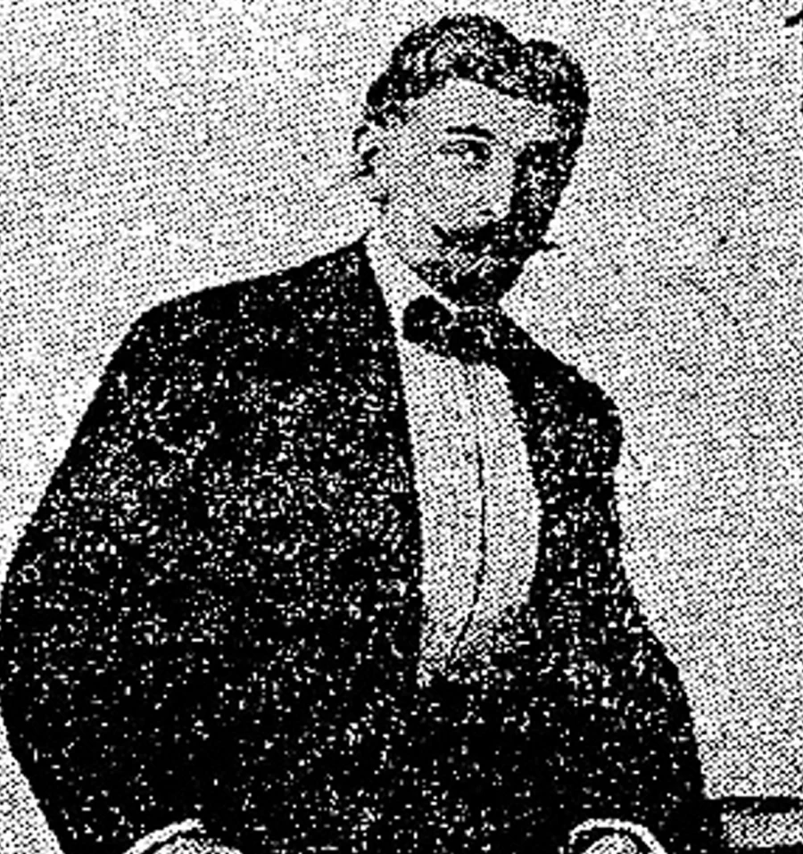
9. Who invented the Van Gieson stain protocol?
Mr. Ira Van Gieson, had the idea of applying unexpected physicochemical principles in histology—principles still used today.

| 470, avenue Laurendeau, Montréal-Est (Quebec) H1B 5M2 | |||
| Phone: | 514 498-3620 | Toll free: | 833 498-3620 |
| Email: | chaptec@chaptec.com | ||
